Laboratory
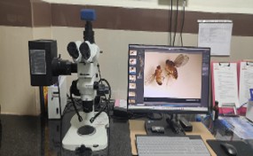
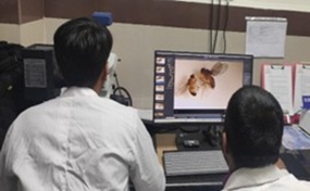
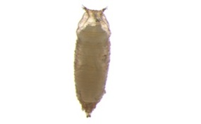
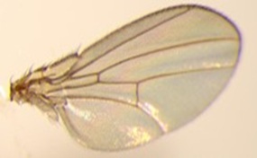
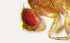
Drosophila lab
Research focus:
Skeletal muscle is central to systemic health; consequently, any disruption to its homeostasis can lead to profound physiological complications. Our laboratory is dedicated to elucidating the complex molecular mechanisms that drive skeletal muscle loss in the context of diabetes. We use Drosophila melanogaster and mouse as model organisms to study the gene regulation in diabetes induced skeletal muscle loss. By exploring how epigenetic modifications influence gene expression in response to metabolic stress, we aim to uncover mechanisms that contribute to the development and progression of diabetes.
Curriculum:
In addition to research, we are deeply committed to training the next generation of geneticists. We lead core modules in Developmental Biology and the utility of Drosophila in biomedical research for both undergraduate and postgraduate Medical Genetics students. The teaching emphasizes the translation of fundamental biological principles from model systems to clinical understanding, fostering a rigorous academic environment that bridges the gap between laboratory discovery and human health.
Cytogenetics lab
The Cytogenetic Laboratory is a specialized diagnostic and research facility dedicated to the study of human chromosomes using samples such as blood, bone marrow, prenatal specimens, and products of conception (POC). The laboratory focuses on identifying chromosomal abnormalities that play a critical role in genetic disorders, infertility, and malignancies.
Equipped with modern infrastructure and adhering to standardized protocols, the laboratory ensures high accuracy, reliability, and quality in all diagnostic services.
Diagnostic Services:
- Peripheral Blood Karyotyping
- Fluorescence In Situ Hybridization (FISH)
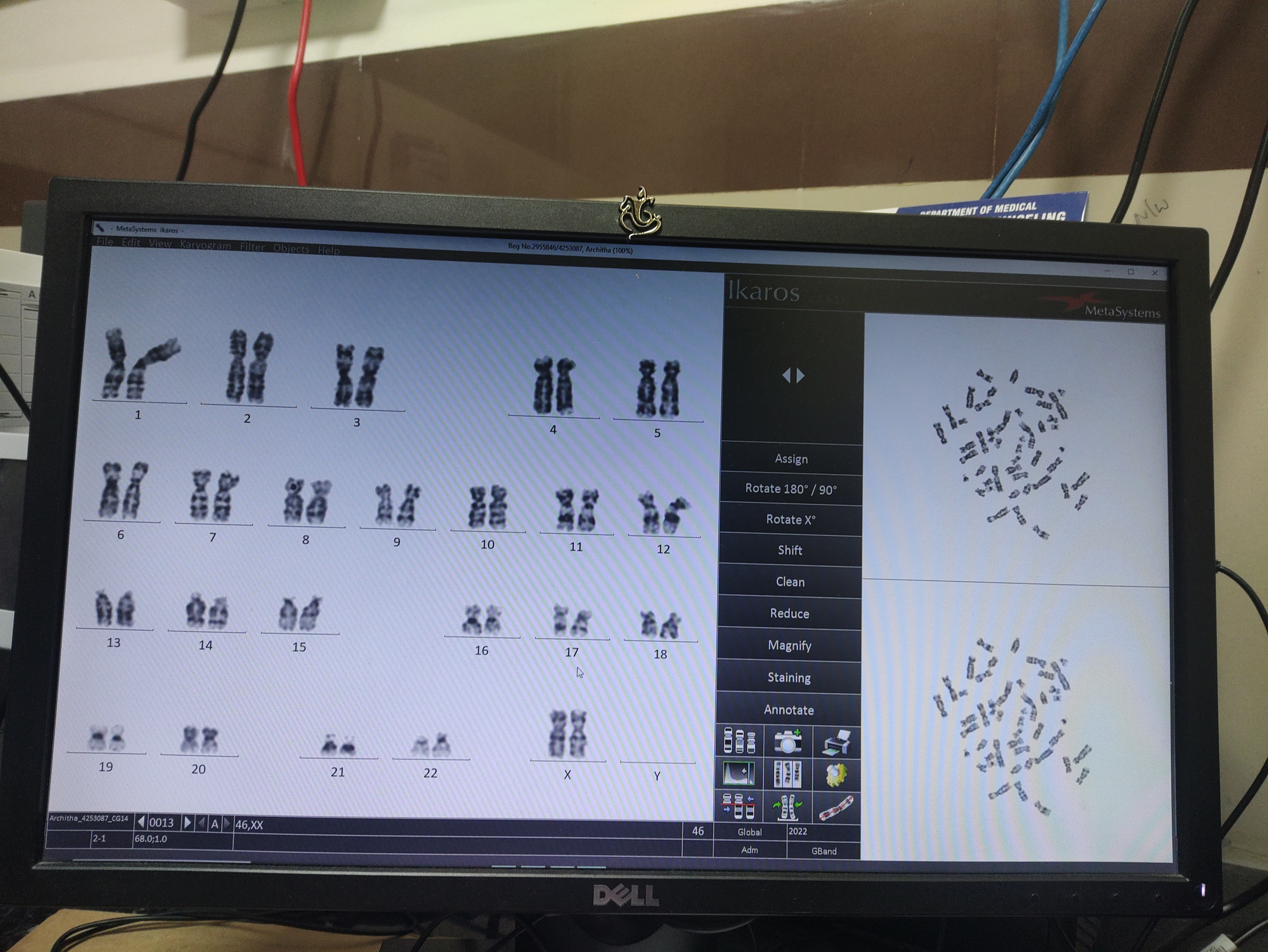
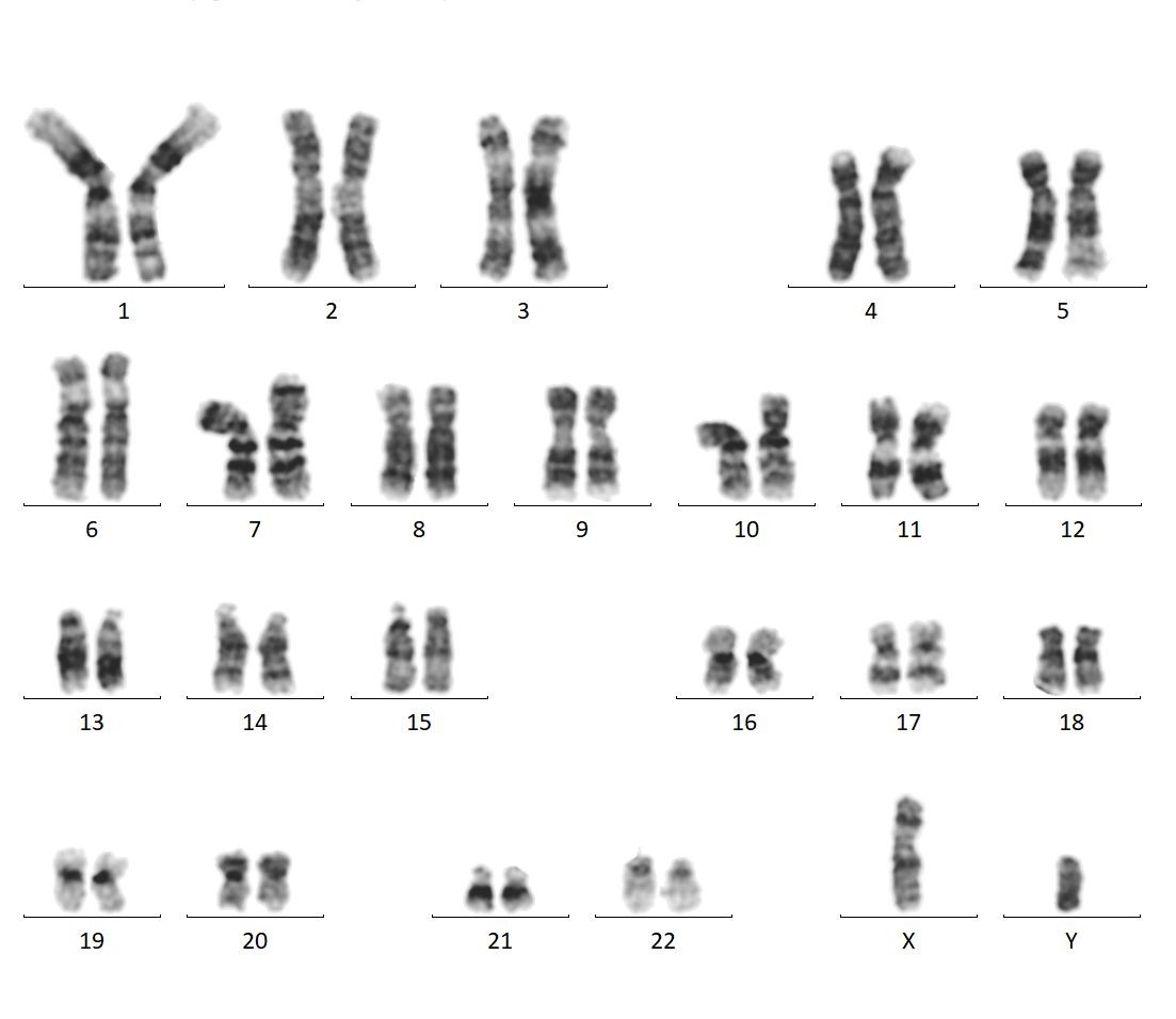
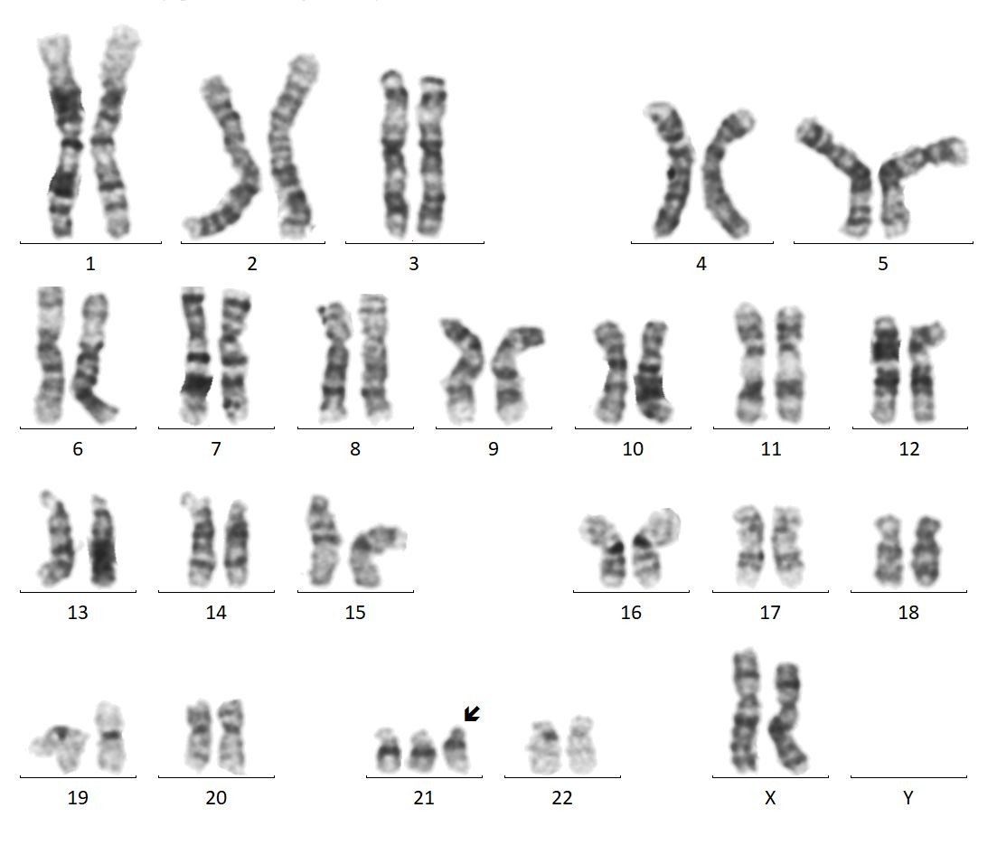
Peripheral Blood Karyotyping
Peripheral Blood Karyotyping is a core service offered by the laboratory. It is a vital cytogenetic technique used to detect both numerical and structural chromosomal abnormalities.
This method enables the identification of:
- Aneuploidies (e.g., trisomies)
- Chromosomal deletions and duplications
- Structural rearrangements and translocations
Indications:
- Congenital anomalies and dysmorphic features
- Developmental delay and intellectual disability
- Disorders of sex development (DSD) / ambiguous genitalia
- Infertility evaluation
- Recurrent pregnancy loss
- Family history of chromosomal abnormalities
- Short stature or pubertal disorders
- Hematological disorders and malignancies
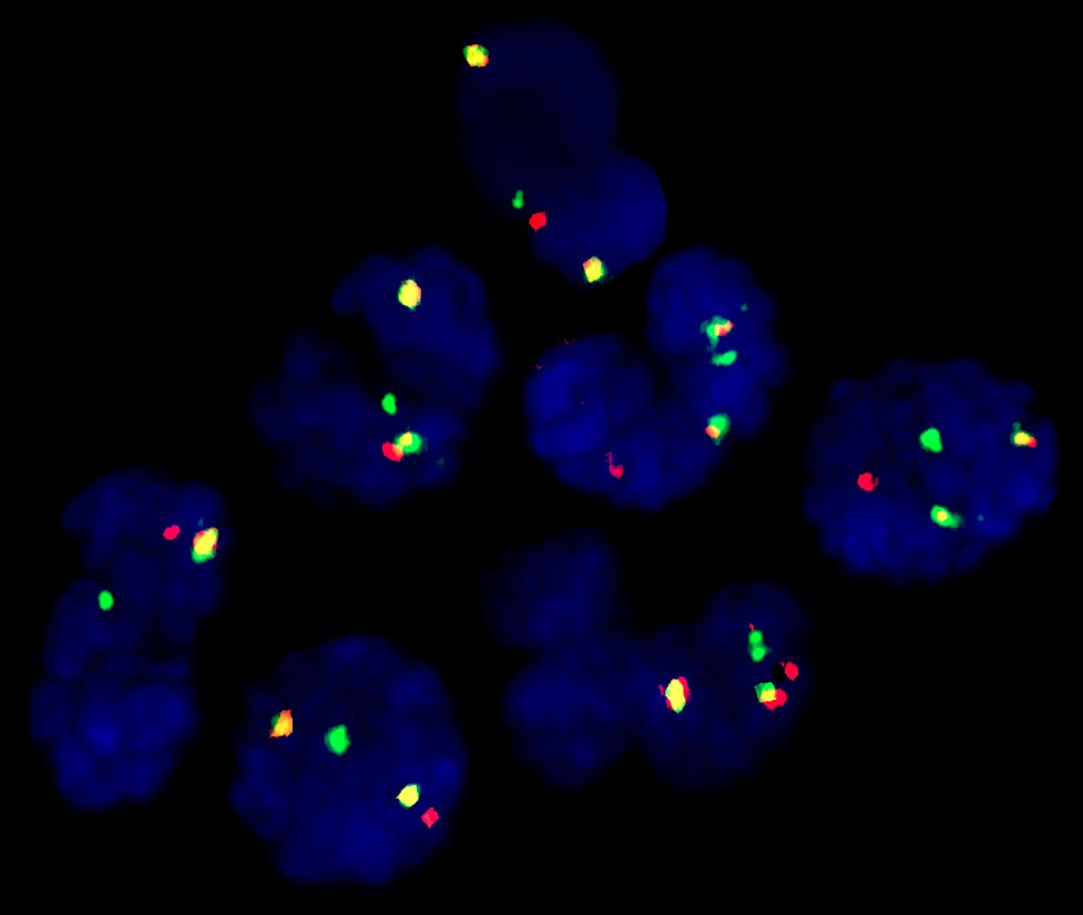
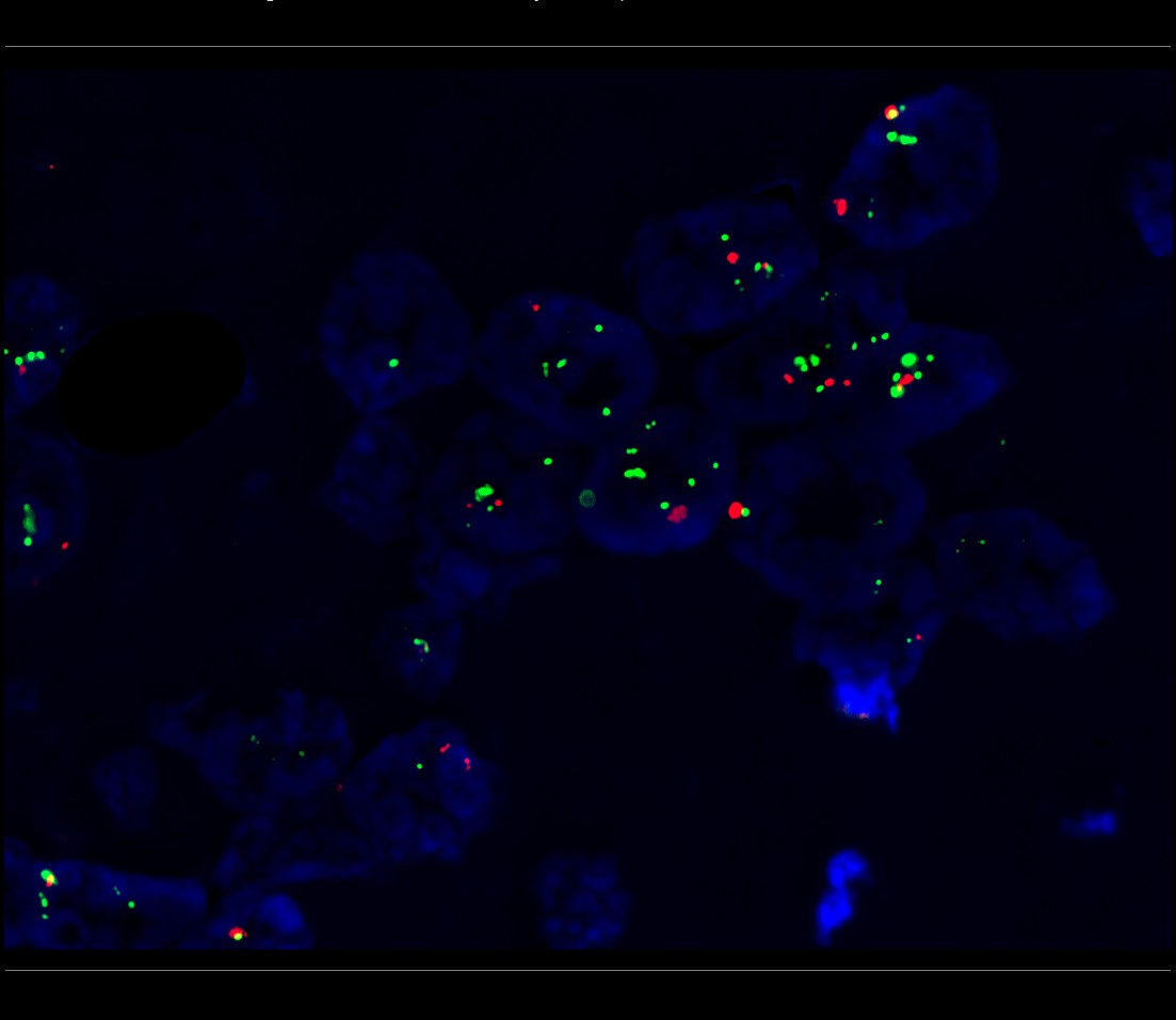
Fluorescence In Situ Hybridization (FISH)
Fluorescence In Situ Hybridization (FISH) is a highly sensitive molecular cytogenetic technique used to detect specific genetic abnormalities at the DNA level. It utilizes fluorescent probes that bind to targeted chromosomal regions, allowing precise visualization under a fluorescence microscope.
Applications:
- Prenatal genetic studies using blood, cord blood, and amniotic fluid
-
Detection of common syndromes such as:
- Down syndrome
- Edward’s syndrome
- Patau syndrome
- Klinefelter syndrome
- Turner syndrome
Specialized FISH Testing:
- BCR/ABL1 FISH for hematological malignancies
- HER2/neu FISH for breast cancer diagnosis and prognosis
Academic and Training Role
In addition to diagnostic services, the Cytogenetic Laboratory serves as an academic and training center for undergraduate and postgraduate students. The facility provides hands-on training and promotes research in the field of human genetics, contributing to the development of future healthcare and research professionals.
Genome lab
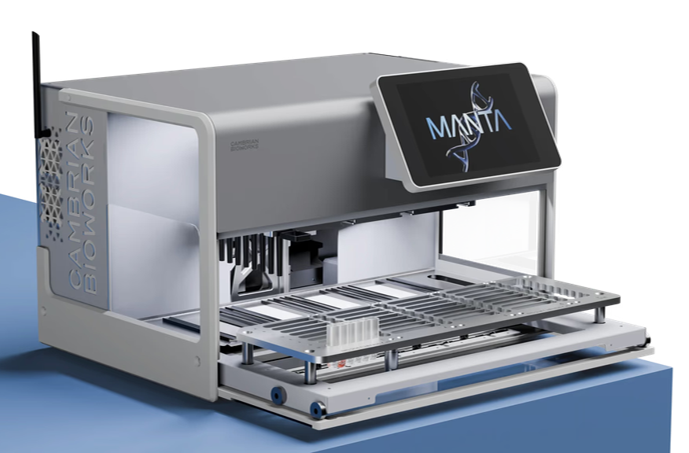
Make : Cambrian Bioworks
Model: Manta
Automated DNA extraction Robot
- Fully automated system for isolation and purification of Nucleic acids from different biological sources
- Generates High quality Nucleic acid preparations for downstream processing NGS, qPCR.
- Enables simultaneous processing of 32 samples
- Reduces hands-on time to under 25-35 minutes for isolation and purification.
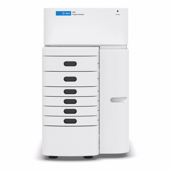
Make : Agilent (Affymetrix)
Model: FA5200
Automated Fragment Analyser — FA5200
- Automated capillary gel electrophoresis system for fragment sizing and quantification.
- Analyses DNA, RNA and proteins with high accuracy and speed in a 96-well format.
- RNA integrity number (RIN) equivalent scoring for RNA quality assessment.
- Used for NGS library quality control, microsatellite analysis and CRISPR editing confirmation.
- Simultaneous processing of up to 96 samples with automated capillary array filling.
- DNF-488 and other dye intercalation kits for broad analyte compatibility.
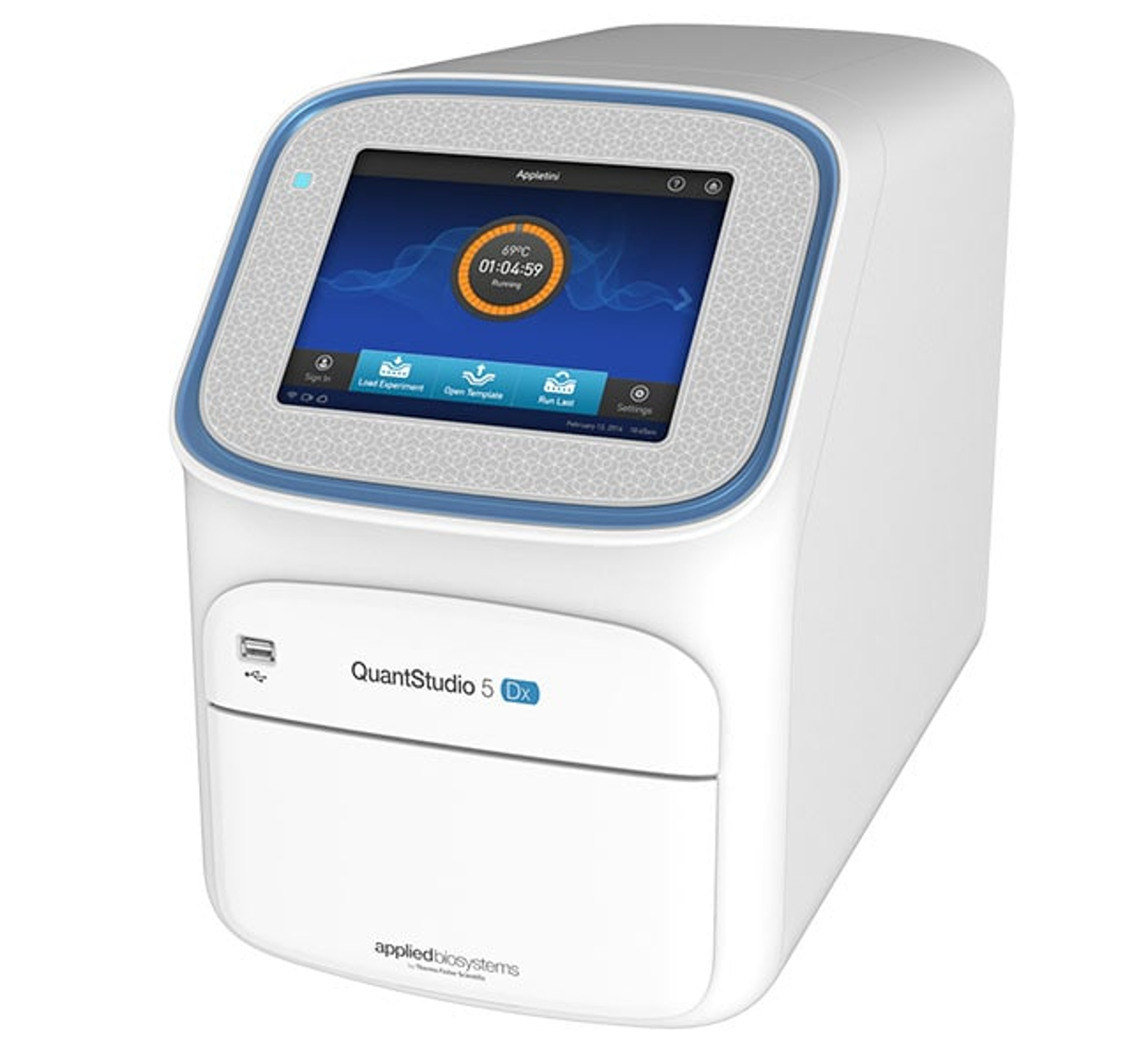
Make : Thermo Fisher Scientific
Model: A32006 – QuantStudio 5 Dx
Real-Time PCR — QuantStudio 5
- 96-well real-time PCR platform with 6-colour optical system for multiplexed detection.
- Compatibility with SYBR Green, TaqMan, High Resolution Melt (HRM) and digital PCR workflows.
- Applied in gene expression, SNP genotyping, copy number variation and viral load quantification.
- IVD-cleared version compliant with CE-IVD and FDA 510(k) for clinical diagnostic use.
- Fast mode cycling completes 40-cycle run in under 40 minutes.
- Connectivity Suite software for GLP compliance, audit trail and 21 CFR Part 11 documentation.
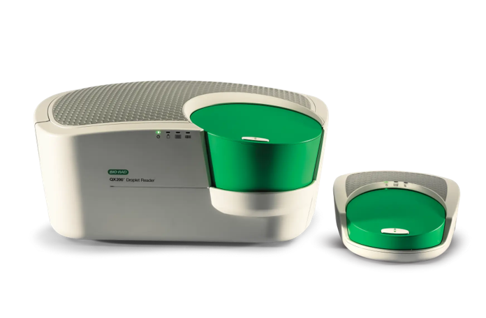
Make : Bio-Rad
Model: QX200 Droplet Reader / Generator
QX200 Droplet Digital PCR
- Digital PCR enabling absolute quantification of nucleic acids without standard curves.
- Partitions samples into 20,000 nanolitre droplets for statistical counting of target copies.
- Sensitivity down to 1 copy in 100,000 wild-type molecules for rare mutation detection.
- Applications: liquid biopsy, ctDNA quantification, GMO detection and viral load measurement.
- Two-channel FAM and HEX/VIC fluorescence detection for duplex assays.
- QX Manager Software with automated cluster calling and CNV analysis modules.
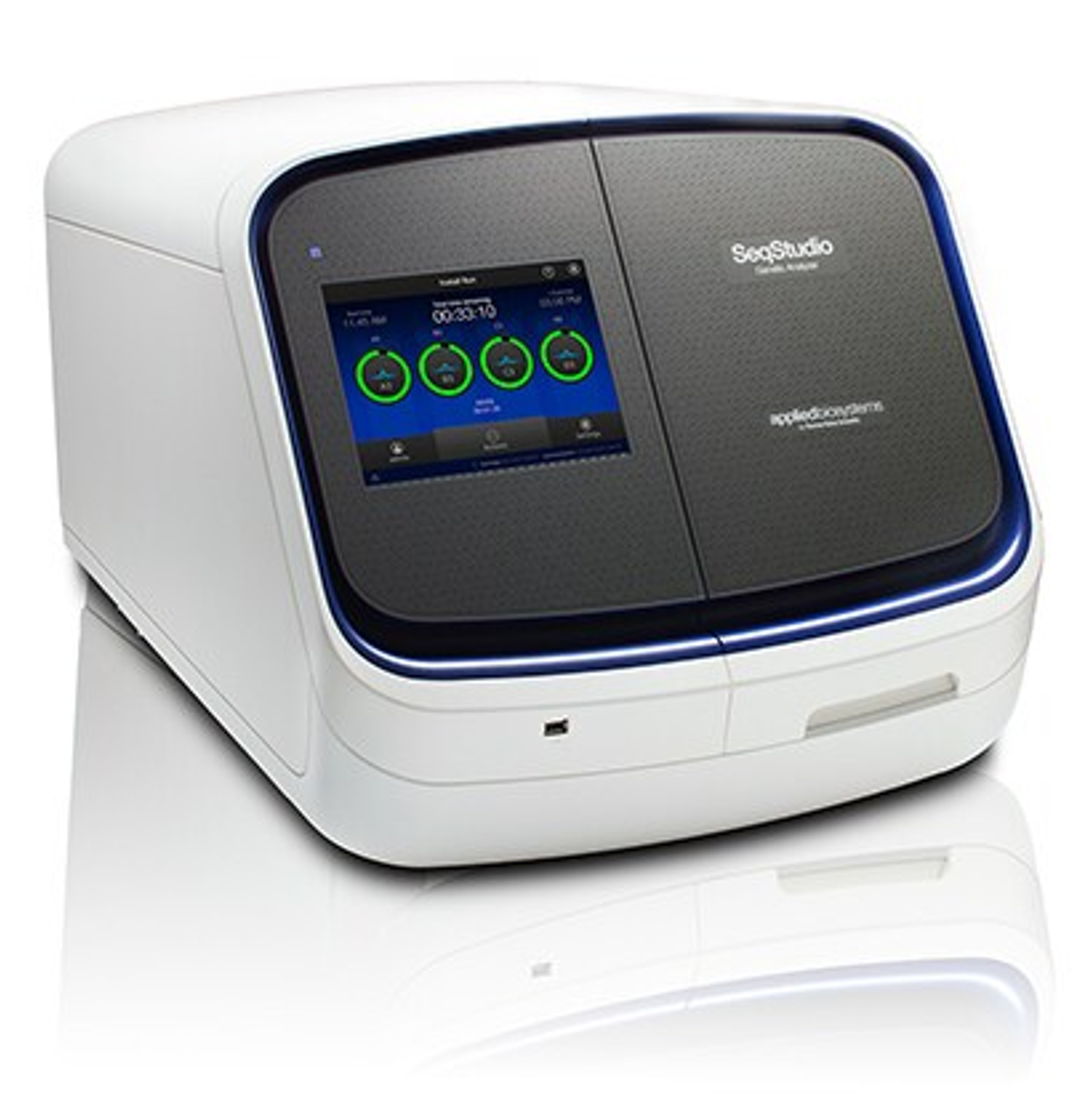
Make : Thermo Fisher Scientific
Model: SeqStudio Genetic Analyser
Capillary Electrophoresis Genetic Analyser — SeqStudio
- Sanger sequencing and fragment analysis on a single 4-capillary instrument.
- Up to four samples run simultaneously with automated polymer and capillary management.
- Dye set F covers BigDye™ Terminator v1.1, v3.1 for sequencing; ROX ladder for fragment analysis.
- Applied in mutation confirmation, STR genotyping, microsatellite instability and LOH analysis.
- Cloud-connected via SeqStudio Data Manager with automatic data backup.
- High-quality sequence data with Phred20 read lengths >900 bases.
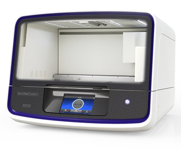
Make : Thermo Fisher Scientific
Model: Ion Chef System
Automated Template Preparation — Ion Chef System
- Fully automated system for Ion Sphere Particle (ISP) preparation for Ion Torrent sequencing.
- Generates clonally-amplified ISPs from purified libraries using one-touch automation.
- Eliminates manual emulsion PCR for consistent template quality run-to-run.
- Supports Ion 520, 530, 540 and 550 chips for variable output requirements.
- Integrated barcode scanner for sample tracking and LIMS connectivity.
- Reduces hands-on time to under 5 minutes from library to loaded chip.
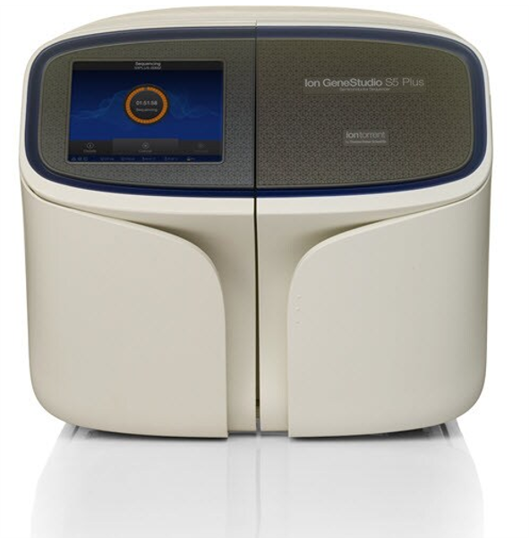
Make : Thermo Fisher Scientific
Model: Ion GeneStudio S5 Plus
Next-Generation Sequencer — Ion GeneStudio S5 Plus
- Semiconductor-based next-generation sequencer for targeted, amplicon and whole-genome sequencing.
- Supports Ion AmpliSeq™ panels for cancer, hereditary disease, pharmacogenomics and microbiome.
- Output range 0.3–15 Gb per run depending on chip selected (Ion 530, 540 or 550 chip).
- Automated library preparation with Ion Chef System for reproducible template preparation.
- On-instrument variant analysis with Torrent Suite software and Variant Caller plugin.
- Ideal for clinical oncology, genetic disease diagnostics and infectious disease surveillance.
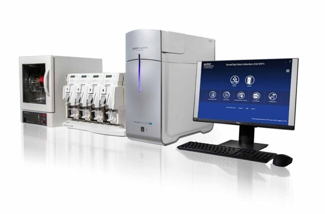
Make : Affymetrix
Model: GeneChip™ System 3000Dx v.2
Microarray Scanner — GeneChip Scanner 3007G
- High-resolution confocal laser scanner for Affymetrix GeneChip microarray analysis.
- Dual-laser excitation (488 nm and 570 nm) for simultaneous two-channel scanning.
- Resolution: 0.7 µm for standard arrays; 5–0.7 µm dynamic range.
- Processes standard, small-format and custom Affymetrix GeneChip arrays.
- Integrated with GeneChip Fluidics Station 450 and Hybridisation Oven 654.
- Applied in gene expression profiling, SNP genotyping, copy number analysis and cytogenetics.
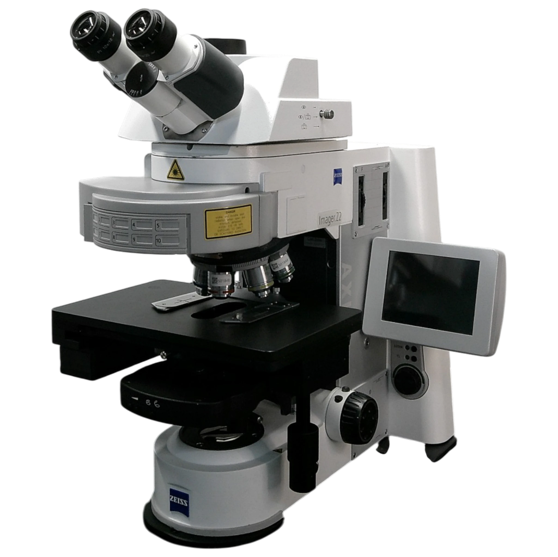
Make : Thermo Fisher Scientific
Model: Ion GeneStudio S5 Plus
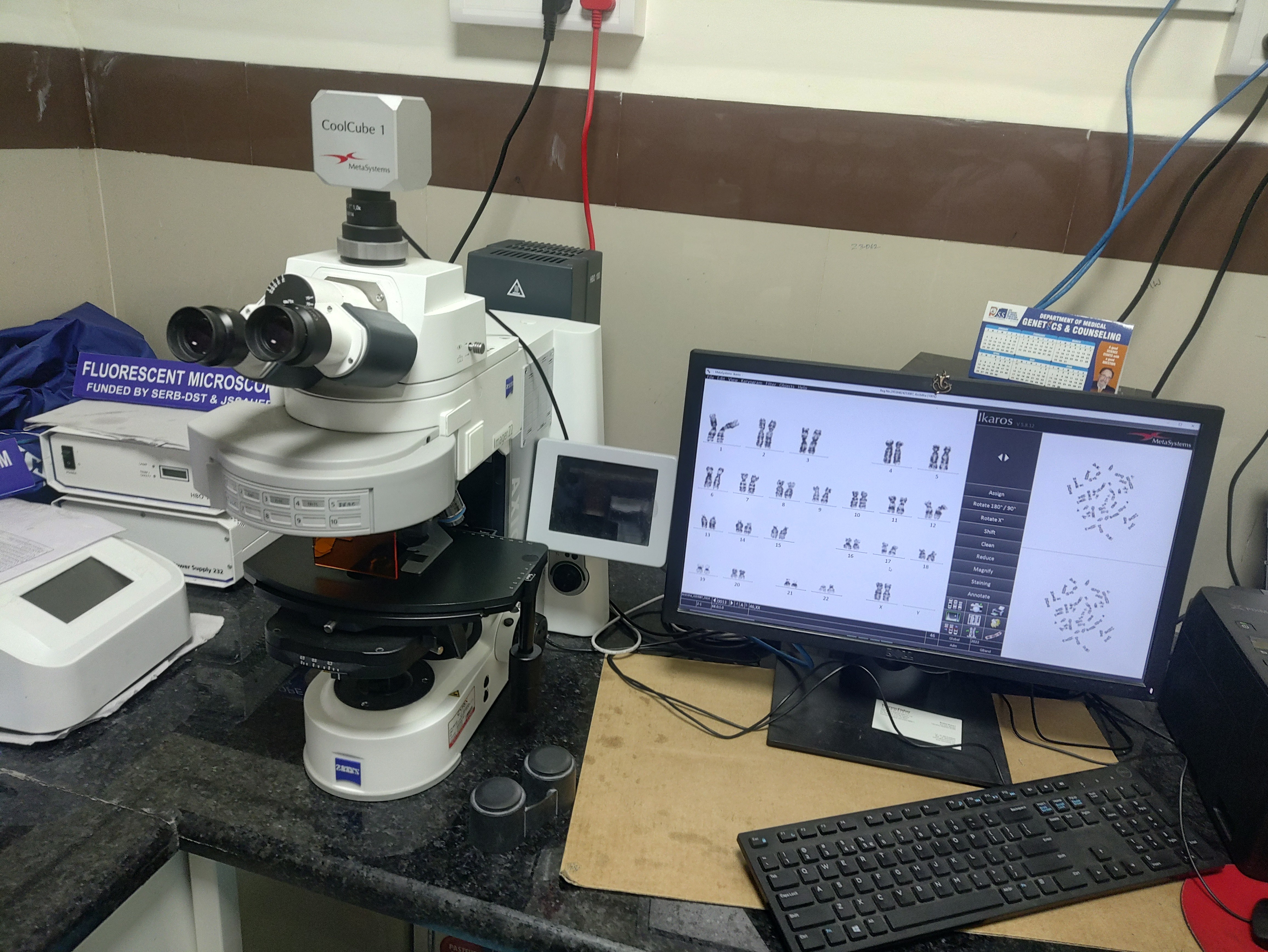
Fluorescence Microscope - Zeiss Axio Imager Z2
- Upright automated fluorescence microscope for karyotyping and cytogenetic analysis.
- Motorised Z-drive with focus reproducibility of less than 0.1 µm for precise Z-stack acquisition.
- Apo-Chromat objectives with high NA for crisp, aberration-free images.
- ISIS karyotyping and FISH analysis software integration for chromosome analysis.
- Multi-band filter sets enabling simultaneous detection of 4+ fluorochromes.
- Applied in prenatal diagnosis, haematological malignancy and gene rearrangement studies.
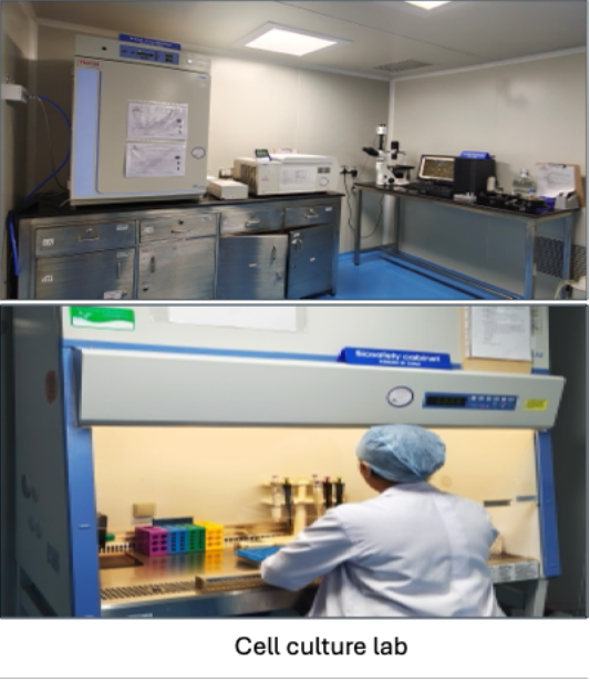
Cell culture lab
Under the blessings of His Holiness and the eminent leadership of the respected Pro-chancellor, JSS Academy of Higher Education and Research has established a state-of Facility to provide research infrastructure.
Our Cell Culture and Organoid Research Laboratory is equipped with advanced infrastructure to support mammalian cell culture, 3D organoid systems, translational research, molecular biology experiments, and preclinical investigations. The facility provides a sterile and controlled environment for routine cell maintenance, disease modelling, drug screening, and cellular characterisation studies.
Applications
- Advanced facility for mammalian cell culture and 3D organoid research
- Supports translational research, molecular biology, and preclinical studies
- Sterile and controlled environment for safe handling of biological samples
- Supports advanced applications in disease modeling, cancer research, regenerative medicine, and personalized therapeutic studies
- Facilitates drug discovery, stem cell–based research, toxicological investigations, and precision healthcare applications
Cell lines maintained
- MDB231
- MCF7
- HepG2
- HuH7
- C2C12
- HCT – Colon adenocarcinoma
- SW48
- SMA Patient derived cell line (
Lab Instrumentation
The laboratory houses state-of-the-art equipment to ensure optimal culture conditions and reliable experimental outcomes, including:
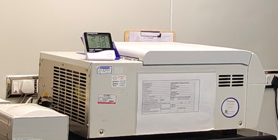
- Thermo Fisher Scientific CO₂ Incubator (Thermo 371): Provides precise control of temperature, humidity, and CO₂ levels for maintaining healthy and contamination-free cell cultures.
- Thermo Fisher Scientific Steri-Cycle CO₂ Incubator: Equipped with advanced contamination control technology to support sensitive cell culture applications and long-term experiments.
- Thermo Fisher Scientific Sorvall Refrigerated Centrifuge: Enables efficient sample processing and cell harvesting while maintaining sample integrity under controlled temperature conditions.
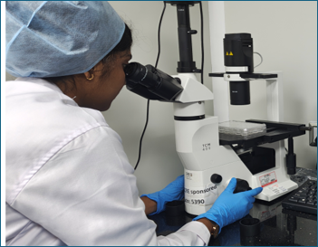
- Inverted Phase Contrast Microscope: Facilitates real-time visualization, monitoring, and assessment of cell morphology, confluency, and organoid growth dynamics.
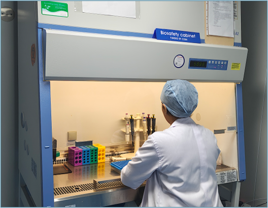
- Thermo Fisher Scientific Class II Biosafety Cabinet (BSL-II): Ensures aseptic handling of cell cultures, organoids, and biological samples while providing operator and environmental protection.
3D Organoid Culture
- In addition to conventional 2D cell culture systems, the laboratory also supports 3D organoid culture technologies, enabling physiologically relevant 3D models facilitating advanced translational research by closely mimicking in vivo tissue architecture and cellular heterogeneity.
- Enables disease modeling, cancer biology studies, regenerative medicine research, and personalized therapy investigations.
- Facilitates drug screening, stem cell research, toxicology studies, and precision medicine applications
The laboratory also serves as a training platform for researchers and students in modern cell culture and organoid methodologies.
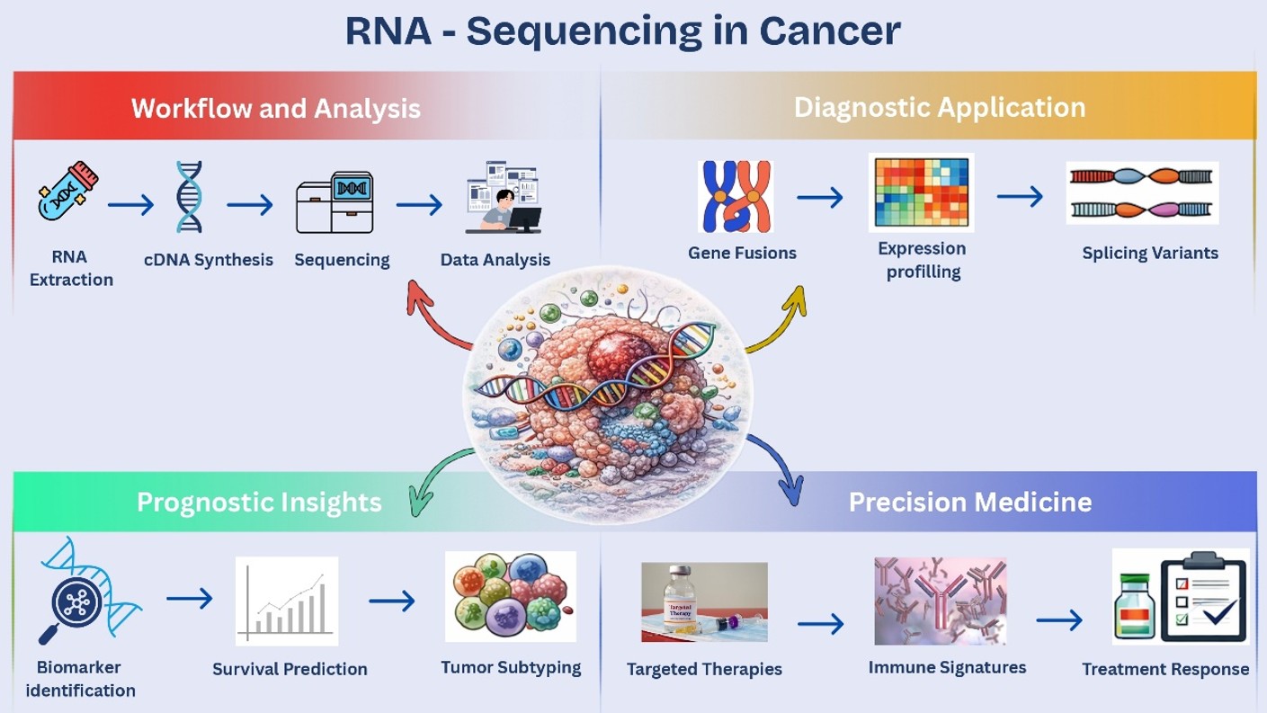
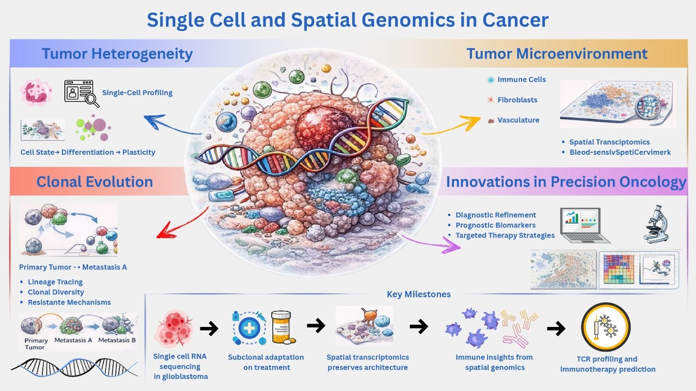
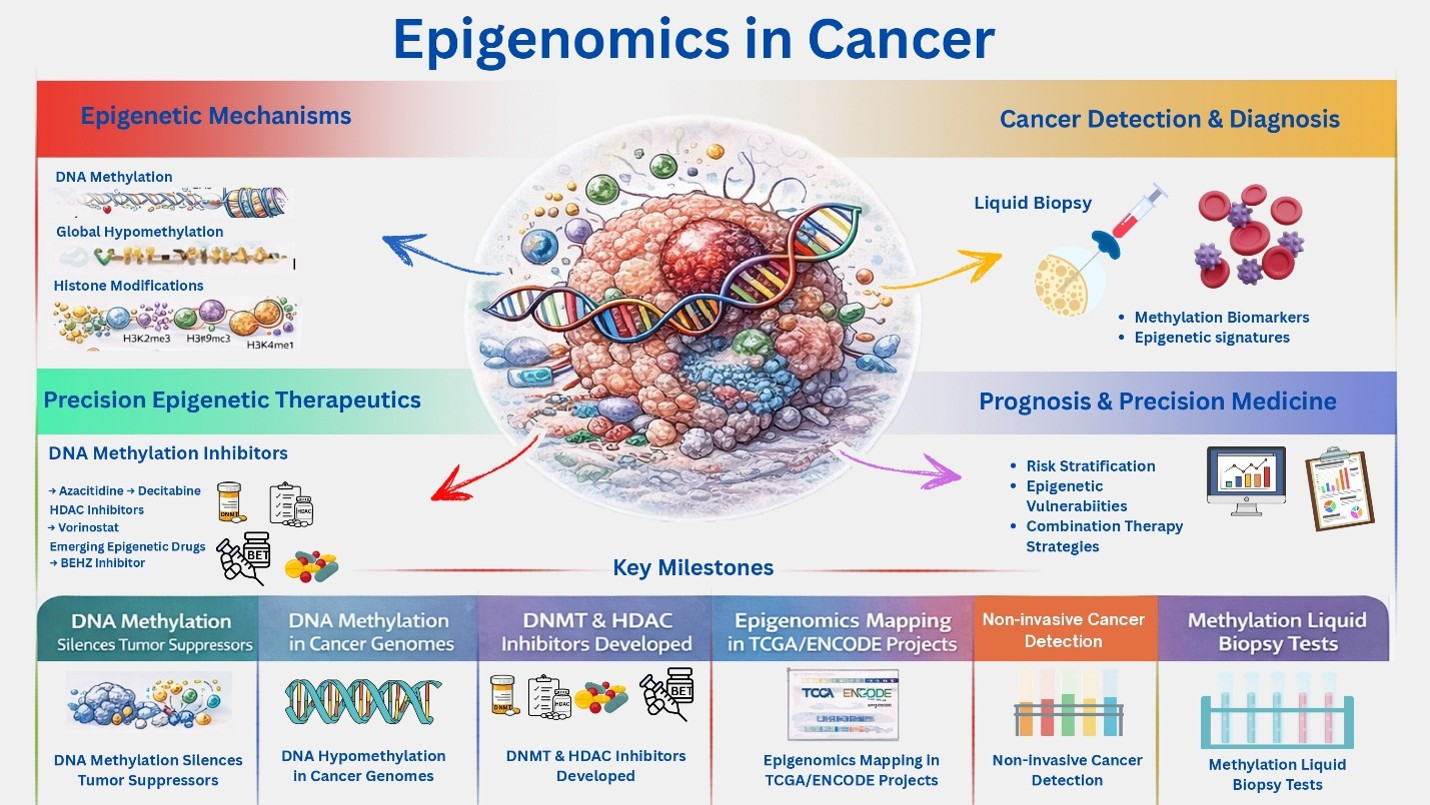
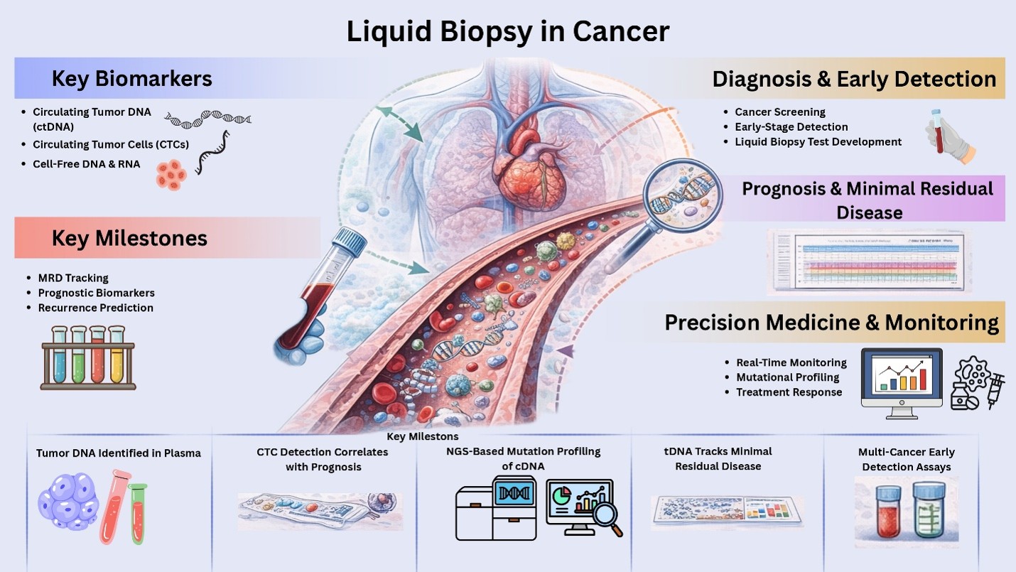
Cancer lab
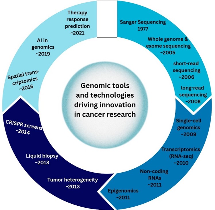
The Cancer Genomics and Translational Oncology Laboratory, led by Dr Suvilesh Kanve Nagaraj, focuses on understanding the molecular and cellular mechanisms driving cancer initiation, progression, metastasis, and therapy resistance. The laboratory integrates disease biology, genomics, cancer informatics, and translational oncology approaches to identify clinically actionable biomarkers and therapeutic targets across multiple cancer types. A major vision of the lab is to bridge basic cancer research with precision medicine by combining patient-derived biospecimens, advanced genomic technologies, bioinformatics, and experimental models. The laboratory employs multidisciplinary approaches, including next-generation sequencing, single-cell omics, computational biology, liquid biopsy analysis, patient-derived organoids and xenografts, genetically engineered mouse models, and molecular and cellular biology techniques, to investigate tumour heterogeneity and tumour-immune interactions.
The lab is particularly interested in deciphering mechanisms underlying metastasis and therapeutic resistance, with the long-term goal of developing improved diagnostic, prognostic, and therapeutic strategies for cancer patients. The lab fosters a highly collaborative and translational research environment by working closely with clinicians, surgeons, pathologists, computational scientists, and basic researchers. In addition to research, the laboratory is committed to mentoring students and young scientists in cancer biology, genomics, and bioinformatics. The research program is supported by intramural and extramural funds awarded to Dr. Suvilesh Kanve Nagaraj. The laboratory actively welcomes academic, clinical, and industrial collaborations in the areas of cancer genomics, precision oncology, and translational cancer research. For collaborations or opportunities to join the lab, please contact Dr. Suvilesh at suvileshkn@jssuni.edu.in